


Recent research published in Nature Neuroscience has overturned long-standing beliefs about how the brain adjusts following the amputation of a limb. For many years, scientists operated under the assumption that the brain’s body map undergoes significant reorganization when a body part is removed, with nearby body parts taking over the space once occupied by the amputated limb.
The new findings reveal that, contrary to this belief, the brain’s body map remains remarkably stable even years after an amputation. This study, led by a team of researchers including Malgorzata Szymanska and Tamar R. Makin, highlights the resilience of the brain’s representation of the body, challenging the foundations of what is known as adult brain plasticity.
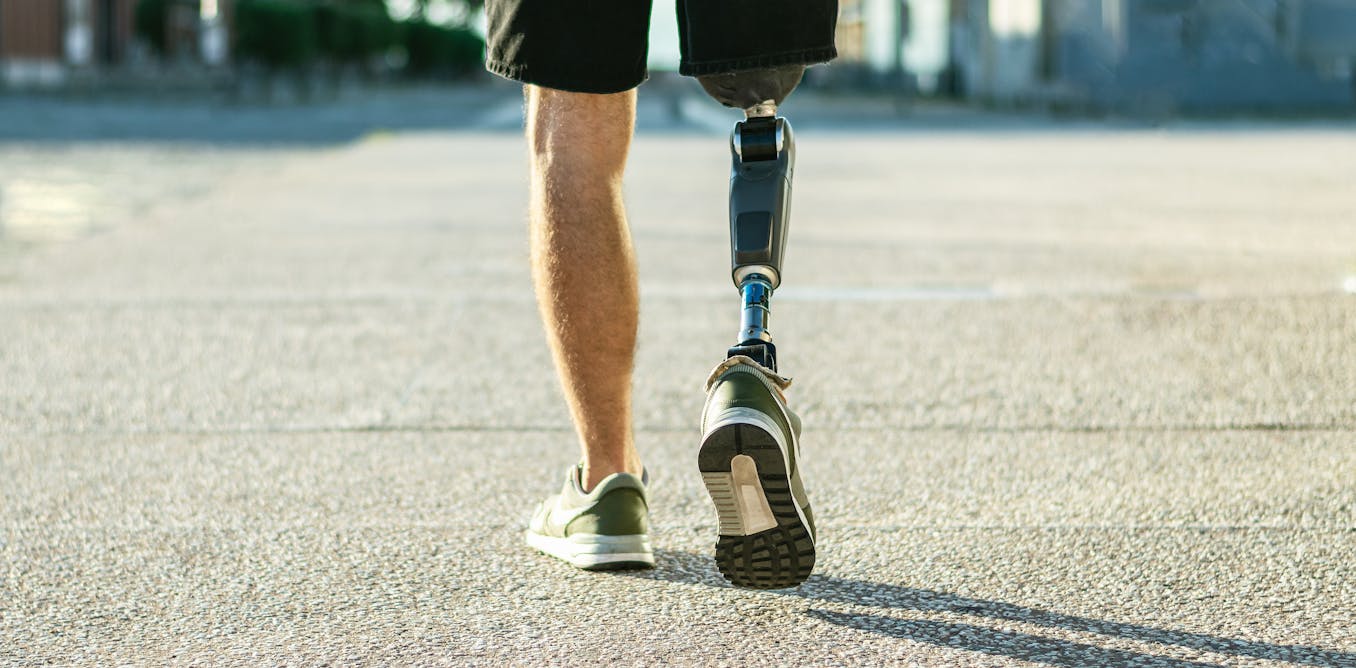
To investigate the brain’s response to amputation, the researchers collaborated with NHS surgeons and conducted functional magnetic resonance imaging (fMRI) scans on three adult patients scheduled for arm amputations due to serious medical conditions. The patients were scanned before their operations and multiple times afterward, in some cases for as long as five years. During these scans, participants were asked to move various body parts, including their fingers and toes, which allowed the researchers to create a detailed map of brain activity associated with each body part.
After the surgeries, the patients were also asked to visualize moving their missing fingers. These “phantom movements” illustrate a phenomenon commonly experienced by amputees, where they continue to feel sensations from their absent limbs. Remarkably, the study found that the brain’s representation of the hand did not change significantly following amputation. Unlike previous theories that suggested brain regions would be taken over by nearby body parts, such as the face, the hand representation remained intact.
This stability of the brain’s body map may help explain why many amputees experience vivid sensations, often described as painful, in their missing limbs. The study suggests that the previous understanding of phantom pain—rooted in the idea of a disorganized brain map—may be misguided. Rather than attempting to fix a supposedly broken map through therapies like mirror box therapy or virtual reality training, which have often proven ineffective, the research indicates that the issue may lie in the severed nerves that misfire signals to the brain.
As a result, new surgical techniques are being developed to preserve nerve signaling during amputations, which could lead to more effective treatments for phantom limb sensations. The implications of this research extend beyond pain management; they also have the potential to advance prosthetics and brain-computer interfaces. Future technologies could leverage the preserved brain map of the amputated limb to enable more intuitive control of artificial limbs, potentially restoring sensations that had been lost.
This innovative study sheds light on the enduring representation of the body within the brain, even in the absence of sensory input. As the researchers conclude, the missing limb continues to exist within the brain, providing both challenges and opportunities for the development of new technologies aimed at improving the lives of amputees.
Malgorzata Szymanska received funding from the Medical Research Council for her PhD and was also supported by the Wellcome Trust while conducting this research. The study benefited from the support of a Wellcome Trust Senior Research Fellowship awarded to Tamar R. Makin. Additionally, Hunter Schone received backing from the Intramural Research Program of the National Institute of Mental Health and a research fellowship from the National Institutes of Health.