


A groundbreaking collaboration has emerged from Melbourne, where scientists successfully studied some of the last preserved Tasmanian tigers in existence. Following extensive negotiations, museum custodians allowed a team to examine the specimens using advanced imaging technology, marking a significant step in understanding this iconic species.
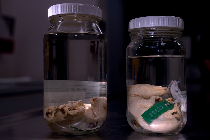
This unprecedented study involved a total of 13 specimens: nine from various museums across Australia and four from Charles University in Prague. The latter had been housed in the institution’s zoology department for over a century but were only recently rediscovered. The Tasmanian Museum and Art Gallery, valuing its decades-old specimens, ensured their safe transport by having a curator personally escort them across the Bass Strait.
The researchers employed computed tomography (CT) scanning, a method traditionally used in medicine for diagnosing injuries and diseases. Instead of dissecting the specimens, the CT scans allowed scientists to visualize the internal structure of the Tasmanian tigers non-invasively. This innovative approach has gained popularity in museums and research institutions, as it enables detailed examinations of specimens while preserving their integrity.
For instance, the scans provided an unexpectedly intricate view of one specimen, a nine-week-old male Tasmanian tiger joey, measuring approximately 20 centimeters. While its developing claws and distinctive stripes were visible externally, the true power of the CT scan lay beneath the surface. Researcher Axel Newton utilized a process called segmentation to identify vital organs, including the heart, lungs, and brain, as well as intricate features like the windpipe and bronchioles.
The scans revealed the young thylacines at five developmental stages, offering insights into how a baby Tasmanian tiger would have matured in its mother’s pouch. Astonishingly, the technology also unveiled discrepancies among the specimens. Two samples, which appeared nearly identical externally, were determined not to be Tasmanian tigers. The scans indicated the presence of additional pelvic bones, suggesting they were likely quolls or Tasmanian devils. This revelation reduced the total count of pouch young Tasmanian tigers from 13 to 11, diminishing the value of the museum’s collection by millions of dollars.
Professor Andrew Pask, an epigeneticist from the University of Melbourne and a key member of the team, expressed enthusiasm for the findings. “It was absolutely magical to peel back and examine all the intricate details visible in the scans,” he remarked during a video call.
CT scanning technology has proven valuable beyond the Tasmanian tiger project. For example, Professor Pask is also investigating the effects of microplastics on male infertility, utilizing similar scanning techniques on embryonic mouse specimens. Additionally, he is working on efforts to potentially revive the thylacine, using CT scans to extract DNA from its teeth.
One of the advantages of CT scanning is that it provides high-resolution, three-dimensional copies of specimens, allowing researchers to study them without risking damage. This approach has garnered interest from museums, as it facilitates educational opportunities. The resulting 3D models can be printed and displayed, enabling the public to engage with the history of this extinct Australian marsupial.
The process of CT scanning involves capturing numerous X-ray images of an object from various angles. These images are then integrated into a digital three-dimensional model, preserving even the most intricate details. At the University of Melbourne, a state-of-the-art micro-CT scanner specializes in imaging small objects, achieving resolutions as fine as 0.001 millimeters.
Dr. Jay Black, a specialist in micro-CT scanning, operates this advanced equipment. He has collaborated across 30 different research fields, utilizing the technology to study everything from meteorites to Tasmanian tiger joeys. The high-resolution capabilities of the micro-CT scanner have proven transformative for researchers like Dr. Jane Melville, a taxonomist who has mapped minute differences between Australian lizard species. Her work has led to the discovery of four distinct species of earless dragons, aiding in conservation efforts for this endangered group.
As the study of museum specimens evolves, researchers are increasingly recognizing the need to democratize access to the vast collections held in institutions worldwide. Dr. Christy Hipsley, another member of the Tasmanian tiger research team, noted that CT scanning could help standardize data sharing, similar to how genomic data is made accessible through databases like GenBank.
In a significant initiative in 2024, numerous natural history museums across the United States collaborated to scan and publish 13,000 vertebrate specimens, showcasing the potential for cross-institutional cooperation in preserving and studying biodiversity.
While the challenges of managing and analyzing extensive datasets persist, researchers are exploring innovative solutions, such as employing machine learning to glean insights from the vast amounts of information generated by CT scans. Dr. Melville is currently engaged in a project aimed at understanding how environmental changes have affected various species, hoping to influence future conservation strategies.
The ongoing advancements in CT scanning illustrate not only the potential for scientific discovery but also the commitment to preserving the legacy of species like the Tasmanian tiger. As the world grapples with the impacts of climate change, these efforts will be crucial in informing strategies to protect biodiversity for generations to come.