


Researchers at Sanford Burnham Prebys, in collaboration with scientists in Hong Kong, have discovered a significant connection between the three-dimensional structure of chromatin and gene regulation. Their findings, published on June 27, 2025, in the journal Genome Biology, provide insights into how the organization of DNA within cells can influence gene activity, with implications for understanding various diseases, including cancer.
Traditionally, the human genome is taught in a linear format, which simplifies its complexity. In reality, DNA is tightly packed into the cell nucleus, resembling a thread wound around protein spools known as histones. This packaged form, referred to as chromatin, exhibits a three-dimensional structure characterized by loops and clumps that may appear random but play a crucial role in genomic function. Structural anomalies in chromatin have been linked to numerous diseases, such as developmental disorders and specific cancers. For instance, nearly 12% of genomic regions in breast cancer cells show chromatin structural issues, and similar problems are associated with T-cell acute lymphoblastic leukemia.
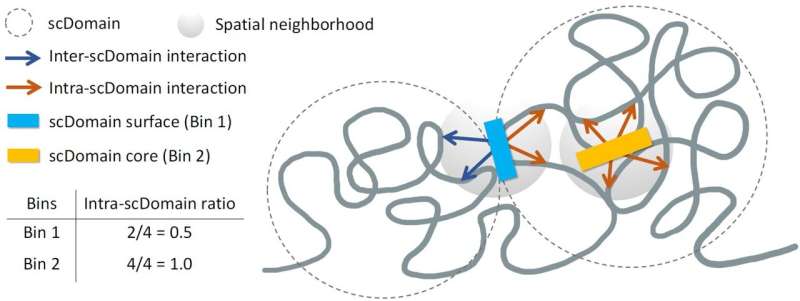
The research team, led by Kelly Yichen Li, Ph.D., a postdoctoral associate at Sanford Burnham Prebys, hypothesized that the 3D shape of these genomic regions profoundly impacts gene regulation. Dr. Li stated, “Many regions of the genome form what are known as topologically associating domains or TADs. Parts of the genome within these domains interact more frequently with each other while remaining isolated from regions outside.”
During their investigation, the researchers employed spatial mapping techniques to analyze the chromatin structures of individual cells. They observed that TAD-like regions often took on globular shapes, with irregularities similar to a selection of potatoes. These shapes led them to suggest that the characteristics of these regions may influence nearby gene functions.
Dr. Yuk-Lap (Kevin) Yip, a professor and interim director of the Center for Data Sciences at Sanford Burnham Prebys, explained, “If you picture these clumps of chromatin fiber being roughly in the shape of a potato, we predicted that regions closer to the surface are more active due to their exposure to biochemical signals in the cell nucleus.” The researchers likened the chromatin’s exterior to the protective skin of a potato, which hinders signals from reaching the densest parts of the chromatin clump.
To validate their hypothesis, the team developed a method to measure the proximity of genomic regions to the core of chromatin clumps. Dr. Li elaborated, “We quantified the ‘coreness’ of a genomic region in a chromatin domain. This measure allowed us to define the surface and core, showing that surface regions are more active than core regions.”
The potential implications of this research are vast. Dr. Yip noted, “The type of data we can apply this measure to is becoming quite plentiful. There is much potential to study how coreness links to gene activity and disease in different cell types.”
Looking ahead, Dr. Li and Dr. Yip plan to collaborate further with Pier Lorenzo Puri, M.D., to deepen their understanding of how the 3D structure of the genome affects muscle stem cell development and the progression of muscular dystrophy. This ongoing research aims to unravel the complexities of gene regulation and its impact on human health.
For further details, refer to the study: Kelly Yichen Li et al, Regulatory roles of three-dimensional structures of chromatin domains, Genome Biology (2025). DOI: 10.1186/s13059-025-03659-7.