


A team of researchers from Washington State University (WSU) has identified a crucial region within a protein known as leiomodin, which is vital for maintaining a healthy heartbeat. This discovery could pave the way for new treatments for serious heart conditions. The findings, published in the journal Circulation Research, highlight the significance of this protein in the regulation of heart muscle function.
Significance of the Discovery
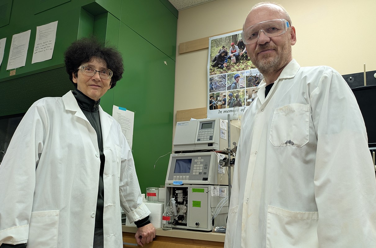
The research reveals that leiomodin plays a critical role in managing the length of thin filaments in heart muscle cells that control heartbeat rhythms. Alla Kostyukova, a professor at the Gene and Linda Voiland School of Chemical Engineering and Bioengineering at WSU, emphasized the importance of this small region of the protein, stating, “It’s a small part of a big protein that turned out to be extremely important for its function in the elongation of thin filaments.”
The heartbeat relies on precise coordination among protein filaments, specifically thick and thin filaments, which respond to electrical signals that enable the heart muscle to contract and relax. The thin filaments are primarily composed of actin, the most abundant protein in the human body. Two additional proteins, tropomodulin and leiomodin, determine the length of these filaments, which is essential for a healthy heart.
Implications for Heart Health
A uniform filament length is crucial for proper heart function at birth and throughout life. For individuals with cardiomyopathy, genetic mutations can result in filaments that are either too short or too long, leading to serious heart issues, including disability and death. Kostyukova noted, “In many cardiomyopathies, the length of the thin filaments is wrong. It always has to be the correct length. If you have a mutation in one of these proteins, your heart cannot work properly.”
Over several years, researchers investigated how leiomodin and tropomodulin compete to regulate filament length. In their recent work, they discovered that a specific region of the leiomodin protein is pivotal for binding to actin and demonstrated its molecular mechanism. Kostyukova explained, “For leiomodin, it’s a weaker binding, and that’s why it was believed that it probably wasn’t binding at all. But we demonstrated that it binds forming a so-called ‘leaky cap,’ allowing it to be removed when the actin starts polymerizing.”
This research utilized nuclear magnetic resonance to analyze the protein’s structure both with and without mutations. Carol Gregorio, a collaborator at the Icahn School of Medicine at Mount Sinai, verified the protein’s behavior in animal cells, supporting the study’s findings.
The team’s collaboration has proven essential in translating structural insights into biological function. Kostyukova remarked, “These proteins are not well known. Now we are going to find out how these binding sites work together in this elongation process. Our hope is to get to the point where we can someday work with small molecules to improve this protein when it has pathogenic mutations.”
Additional contributors to the study from WSU include Garry Smith and Dmitri Tolkachev, an assistant professor in the Voiland School. The research received funding from the National Institutes of Health and the American Heart Association.