


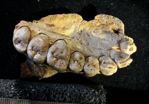
Research has revealed potential risks associated with a commonly used technique for examining fossils, suggesting that micro-computerised tomography (micro-CT) might inadvertently damage these invaluable specimens. Despite its reputation as a non-destructive method, findings published in the journal Radiocarbon indicate that micro-CT can significantly alter the internal structure of fossils, potentially erasing crucial information.
Fossils serve as critical records of Earth’s biological history, offering insights into the evolution of species and past environments. They provide us with a glimpse into ancient diets and migration patterns, including those of early humans. However, studying these fragile remnants poses challenges, as scientists strive to balance the need for research with the imperative to preserve these rare specimens for future generations.
Micro-CT operates similarly to medical CT scans, enabling researchers to create high-resolution three-dimensional images without physically damaging the fossils. This capability allows scientists to explore the internal architecture of fossil specimens while keeping them intact. The technology has been instrumental in various studies, including investigations into the earliest indications of bone cancer in humans and the examination of brain structures in early hominins.
Nevertheless, the recent study led by Mathieu Duval and Laura Martín-Francés uncovered that micro-CT scanning could alter the collagen content in fossils. Collagen is a vital component for numerous analytical techniques, such as radiocarbon dating and stable isotope analysis, which are used to determine the age and diet of extinct species.
While the researchers confirmed that the radiocarbon dating remained unaffected after scanning, they observed a concerning reduction in collagen levels. The scanned samples exhibited approximately 35% less collagen compared to unscanned counterparts, indicating that micro-CT may compromise the integrity of fossils that retain collagen traces.
The study highlights that the exposure to micro-CT’s X-rays, which are a form of ionising radiation, may have more significant effects than previously understood. Although medical applications of X-rays are carefully monitored to minimize risk, the implications for fossils have not been thoroughly investigated until now.
In earlier research, the team found that micro-CT could artificially age fossils analyzed with electron spin resonance, a dating method for specimens over 50,000 years old. This previous discovery, combined with the current findings, suggests that micro-CT scanning could lead to irreversible changes in fossils and the valuable data they hold.
Given these insights, the authors of the study recommend that scientists exercise caution when using micro-CT, advocating for its sparing application to limit exposure to X-rays. They also emphasize the importance of sharing data to prevent unnecessary repeated scans of the same specimens.
The implications of this research extend beyond individual fossils, posing questions about the broader methodologies employed in paleontological studies. With fossils being irreplaceable treasures of our planet’s history, understanding how to protect them while still advancing scientific knowledge is essential.
The study received funding from the Spanish State Research Agency and forms part of ongoing research initiatives under the HORIZON-MSCA-2021-PF-01 program. As the scientific community continues to refine its techniques, the findings serve as a crucial reminder of the inherent fragility of fossils and the need to adapt methodologies to safeguard these critical links to our past.