


Researchers at the Barcelona Institute of Science and Technology (BIST) have achieved a groundbreaking milestone in reproductive biology by filming the exact moment a human embryo implants into the uterine lining. This significant accomplishment opens new avenues for understanding early human development and could enhance fertility treatments. The study, published in Science Advances, marks the first time the implantation process has been dynamically captured on film.
Embryo implantation is a critical step that typically occurs five to six days after fertilization, when a cluster of 100 to 200 cells seeks to attach itself to the uterine wall. Until now, scientists had only been able to take snapshots of this phenomenon, as embryos are too small to be observed with ultrasound at this early stage. This new approach allows for continuous observation, providing insights into one of the most crucial stages of human development.
New Technology Reveals Implantation Process
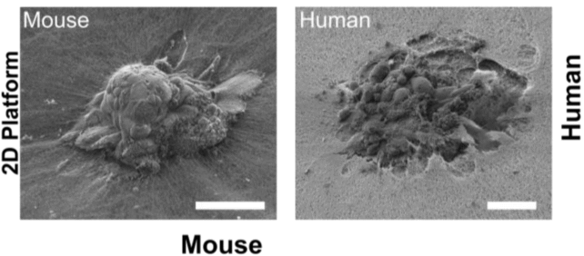
The innovative system developed by senior author and bioengineer Samuel Ojosnegros and his team recreates the structural environment necessary for embryos to implant. During the experiments, researchers observed human embryos penetrating a collagen-based matrix, creating a cavity for further growth. This process was markedly different from that of mouse embryos, which only superficially invaded the matrix. Ojosnegros noted, “Our technology lets us pinpoint where the embryo exerts force, and we found it applies significant mechanical force to implant and invade.”
This finding emphasizes the limitations of relying solely on mouse studies to understand human implantation. Approximately 60 percent of failed pregnancies occur during implantation or shortly after, making this stage a vital area of study for improving reproductive outcomes.
The new uterine model not only extends observation beyond the initial days of embryo development but can also be utilized in both two-dimensional and three-dimensional formats. When placed on a flat gel, embryos could be seen binding to the collagen surface and later invading it. In contrast, when situated within droplets, embryos appeared to ‘pull’ collagen fibers toward themselves, suggesting a complex interaction with their environment.
Implications for Fertility Treatments
Lead author Amélie Luise Godeau and her research team speculate that this interaction may play a role in how embryos connect with maternal tissues. Although the collagen matrix used in the study does not incorporate human uterine cells, it offers a unique opportunity to manipulate its composition to observe how embryos respond to various conditions. This could lead to identifying factors that enhance implantation rates.
Ojosnegros highlighted that their spin-off company, Serabiotics, in collaboration with the pharmaceutical company Grifols, has already developed a protein supplement aimed at improving implantation rates in clinical settings. This advancement showcases the potential real-world applications of their research.
The ability to film and study implantation could provide crucial insights into the early stages of human life, potentially leading to improved fertility treatments and better outcomes for those experiencing difficulties in conceiving. The research not only expands our understanding of human development but also holds promise for future advancements in reproductive health.