


Researchers at the Barcelona Institute of Science and Technology have achieved a groundbreaking milestone in reproductive science by capturing the moment of human embryo implantation for the first time. This significant advancement provides insights into early human development and could lead to improved fertility treatments. The process of implantation, which occurs deep within the uterus, is crucial for establishing a pregnancy, yet it has remained largely mysterious until now.
Using a novel system, scientists filmed human embryos as they penetrated a collagen-based matrix, creating a cavity essential for their connection to the maternal environment. The time-lapse recordings reveal how these embryos aggressively invade the matrix, marking a dynamic and invasive process critical for successful implantation. According to senior author and bioengineer Samuel Ojosnegros, “For the first time, we’ve been able to watch human embryo implantation unfold dynamically. We’ve opened a window into a stage of development that was previously hidden.”
Innovative Techniques Illuminate Development
The experiments were conducted in a laboratory setting, distinct from a natural uterus, yet the platform developed by Ojosnegros and his colleagues effectively simulates the necessary structural and nutritional environment for embryo implantation. This is particularly important because approximately 60 percent of failed pregnancies occur during the implantation phase or shortly thereafter.
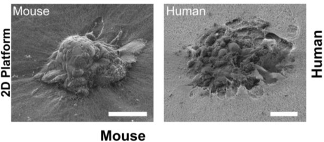
Compared to mouse embryos, which tend to invade the matrix superficially, human embryos demonstrated a more profound invasion, fully enveloping themselves in the collagen material. Ojosnegros noted, “Our technology lets us pinpoint where the embryo exerts force, and we found it applies significant mechanical force to implant and invade. This means that mouse studies can only take us so far in understanding human implantation.”
Typically, a human embryo implants around five to six days after fertilization, when it consists of a cluster of 100 to 200 cells—too small to be detected by ultrasound. The new uterine model extends the observation window for embryos beyond the initial five days, allowing for more comprehensive studies.
Future Implications for Fertility Treatments
The technology can be utilized in various forms, including a flat gel or droplet, to observe embryo implantation in both 2D and 3D. When blastocysts are placed on a flat gel, they can be seen binding to the collagen surface before invading it. In contrast, when embryos are placed within droplets, they appear to ‘pull’ collagen fibers toward their center, reshaping the surrounding environment.
Lead author Amélie Luise Godeau expressed the potential for this research to connect the maternal environment with embryonic tissues. While the study does not explore how the uterine wall responds to this connection, it opens avenues for further inquiry.
The collagen-based matrix used in the study lacks human uterine cells, providing a partial view of the implantation process. Nevertheless, this limitation enables researchers to modify the matrix composition to examine how human embryos respond to different conditions or compounds that could enhance implantation success. Ojosnegros, who co-founded the spin-off Serabiotics, mentioned that they are collaborating with the pharmaceutical company Grifols to develop a protein supplement aimed at increasing implantation rates in clinical settings.
This pioneering study was published in Science Advances, marking a significant step forward in understanding early human development and enhancing reproductive health. The implications of this research could resonate across the fields of fertility and developmental biology, providing hope to many facing challenges in conception.